Datei:Free-living amebic infections.png
Seiteninhalte werden in anderen Sprachen nicht unterstützt.
Aus Wikibooks

Originaldatei zum Herunterladen(1.365 × 1.170 Pixel, Dateigröße: 715 KB, MIME-Typ: image/png)

Dieses Medium wird direkt von Wikimedia Commons aus eingebunden. Quellenangaben und Lizenzbedingungen befinden sich auf der unten zusätzlich eingeblendeten Commons-Beschreibungsseite.

Zur Commons-Seite

Dieses Bild des Typs Biology sollte als Vektorgrafik im SVG-Format neu erstellt werden. Vektorformate haben zahlreiche Vorteile; weitere Information unter Commons:Media for cleanup. Wenn dir eine SVG-Version dieses Bildes vorliegt, so lade diese bitte hoch. Nach dem Hochladen der Datei ist diese Vorlage auf der aktuellen Bildbeschreibungsseite durch die Vorlage {{Vector version available}}, oder kürzer {{Vva}}, zu ersetzen. Es ist empfohlen die neue SVG-Datei „Free-living amebic infections.svg“ zu nennen – dann benötigt die Vorlage vector version available (bzw. vva) keinen Parameter.

Beschreibung

Beschreibung
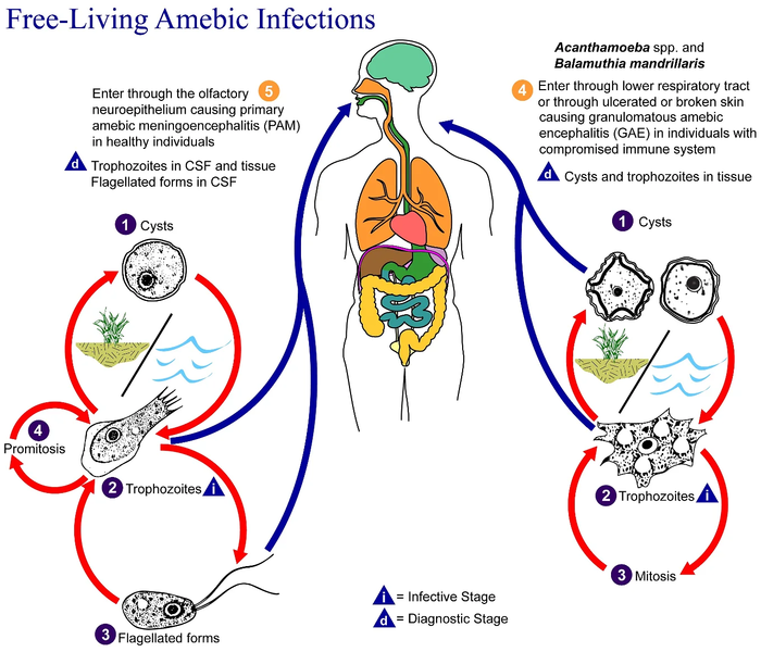
English: This is an illustration of the life cycle of the parasitic agents responsible for causing “free-living” amebic infections.

For a complete description of the life cycle of these parasites, select the link below the image or paste the following address in your address bar: http://www.dpd.cdc.gov/dpdx/HTML/FreeLivingAmebic.htm Free-living amebae belonging to the genera Acanthamoeba, Balamuthia, and Naegleria are important causes of disease in humans and animals. Naegleria fowleri produces an acute, and usually lethal, central nervous system (CNS) disease called primary amebic meningoencephalitis (PAM). N. fowleri has three stages, cysts (1) , trophozoites (2) , and flagellated forms (3) , in its life cycle. The trophozoites replicate by promitosis (nuclear membrane remains intact) (4) . Naegleria fowleri is found in fresh water, soil, thermal discharges of power plants, heated swimming pools, hydrotherapy and medicinal pools, aquariums, and sewage. Trophozoites can turn into temporary flagellated forms which usually revert back to the trophozoite stage. Trophozoites infect humans or animals by entering the olfactory neuroepithelium (5) and reaching the brain. N. fowleri trophozoites are found in cerebrospinal fluid (CSF) and tissue, while flagellated forms are found in CSF.

Acanthamoeba spp. and Balamuthia mandrillaris are opportunistic free-living amebae capable of causing granulomatous amebic encephalitis (GAE) in individuals with compromised immune systems. Acanthamoeba spp. have been found in soil; fresh, brackish, and sea water; sewage; swimming pools; contact lens equipment; medicinal pools; dental treatment units; dialysis machines; heating, ventilating, and air conditioning systems; mammalian cell cultures; vegetables; human nostrils and throats; and human and animal brain, skin, and lung tissues. B. mandrillaris however, has not been isolated from the environment but has been isolated from autopsy specimens of infected humans and animals. Unlike N. fowleri, Acanthamoeba and Balamuthia have only two stages, cysts (1) and trophozoites (2) , in their life cycle. No flagellated stage exists as part of the life cycle. The trophozoites replicate by mitosis (nuclear membrane does not remain intact) (3) . The trophozoites are the infective forms and are believed to gain entry into the body through the lower respiratory tract, ulcerated or broken skin and invade the central nervous system by hematogenous dissemination (4). Acanthamoeba spp. and Balamuthia mandrillaris cysts and trophozoites are found in tissue.
Datum
Quelle

Dieses Medium stammt aus der Public Health Image Library (PHIL), mit der Identifikationsnummer #3412 der Centers for Disease Control and Prevention.

Hinweis: Nicht alle PHIL-Bilder sind gemeinfrei; überprüfe unbedingt den Urheberrechtsstatus und die Nennung der Autoren und Inhaltsanbieter.


العربية | Deutsch | English | македонски | slovenščina | +/−

Urheber
  • Photo Credit:
  • Content Providers(s): CDC/Alexander J. da Silva, PhD/Melanie Moser
Genehmigung
(Weiternutzung dieser Datei)
English: None - This image is in the public domain and thus free of any copyright restrictions. As a matter of courtesy we request that the content provider be credited and notified in any public or private usage of this image.
Andere Versionen

Lizenz

Public domain
Dieses Bild ist ein Werk der Centers for Disease Control and Prevention, einer dem Gesundheitsministerium der Vereinigten Staaten unterstellten Behörde, oder es wurde von einem Mitarbeiter dieser Behörde in Ausübung seiner dienstlichen Pflichten erstellt. Als ein Werk der US-amerikanischen Bundesregierung ist dieses Werk in den Vereinigten Staaten gemeinfrei.

eesti  Deutsch  čeština  español  português  English  français  Nederlands  polski  slovenščina  suomi  македонски  українська  日本語  中文(简体)‎  中文(繁體)‎  العربية  +/−

Ursprüngliches Datei-Logbuch

Übertragen aus en.wikipedia nach Commons durch Optigan13.

Die ursprüngliche Dateibeschreibungsseite war hier. Alle folgenden Benutzernamen beziehen sich auf en.wikipedia.
  • 2006-05-04 01:09 Keenan Pepper 518×435×8 (31658 bytes) Free-living_amebic_infections.gif, converted to [[PNG]] by [[netpbm]].

Kurzbeschreibungen

Ergänze eine einzeilige Erklärung, was diese Datei darstellt.

In dieser Datei abgebildete Objekte

Motiv

image/png

Dateiversionen

Klicke auf einen Zeitpunkt, um diese Version zu laden.

Version vomVorschaubildMaßeBenutzerKommentar
aktuell11:24, 2. Feb. 2023Vorschaubild der Version vom 11:24, 2. Feb. 20231.365 × 1.170 (715 KB)Materialscientisthttps://answersingenesis.org/biology/microbiology/the-genesis-of-brain-eating-amoeba/
08:30, 20. Jul. 2008Vorschaubild der Version vom 08:30, 20. Jul. 2008518 × 435 (31 KB)Optigan13{{Information |Description={{en|This is an illustration of the life cycle of the parasitic agents responsible for causing “free-living” amebic infections. For a complete description of the life cycle of these parasites, select the link below the image

Die folgende Seite verwendet diese Datei:

Globale Dateiverwendung

Die nachfolgenden anderen Wikis verwenden diese Datei:

Metadaten